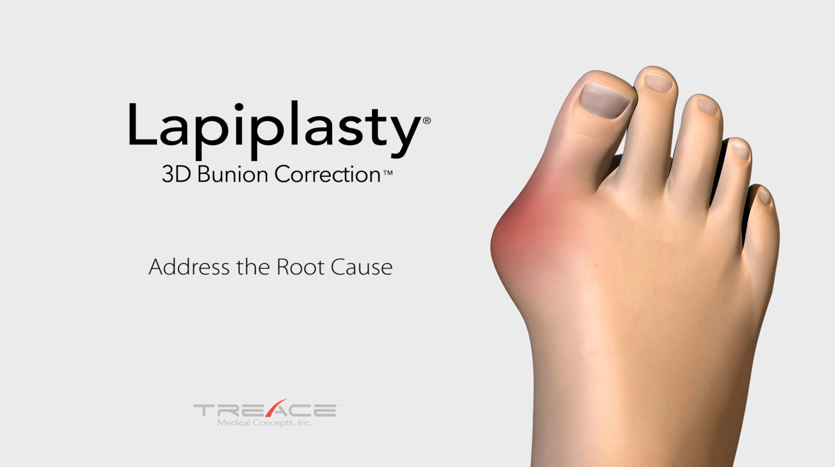
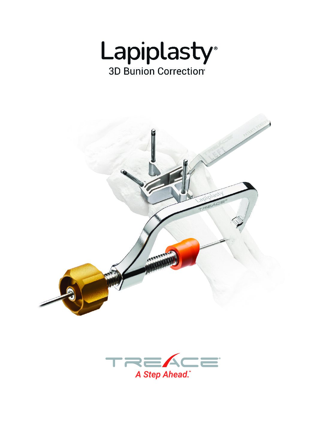
Lapiplasty® 3-Plane Correction at the CORA
- Instrumented Reproducibility
- Rapid Weight-Bearing1
- Low Recurrence at 13-17 month follow-up1,2
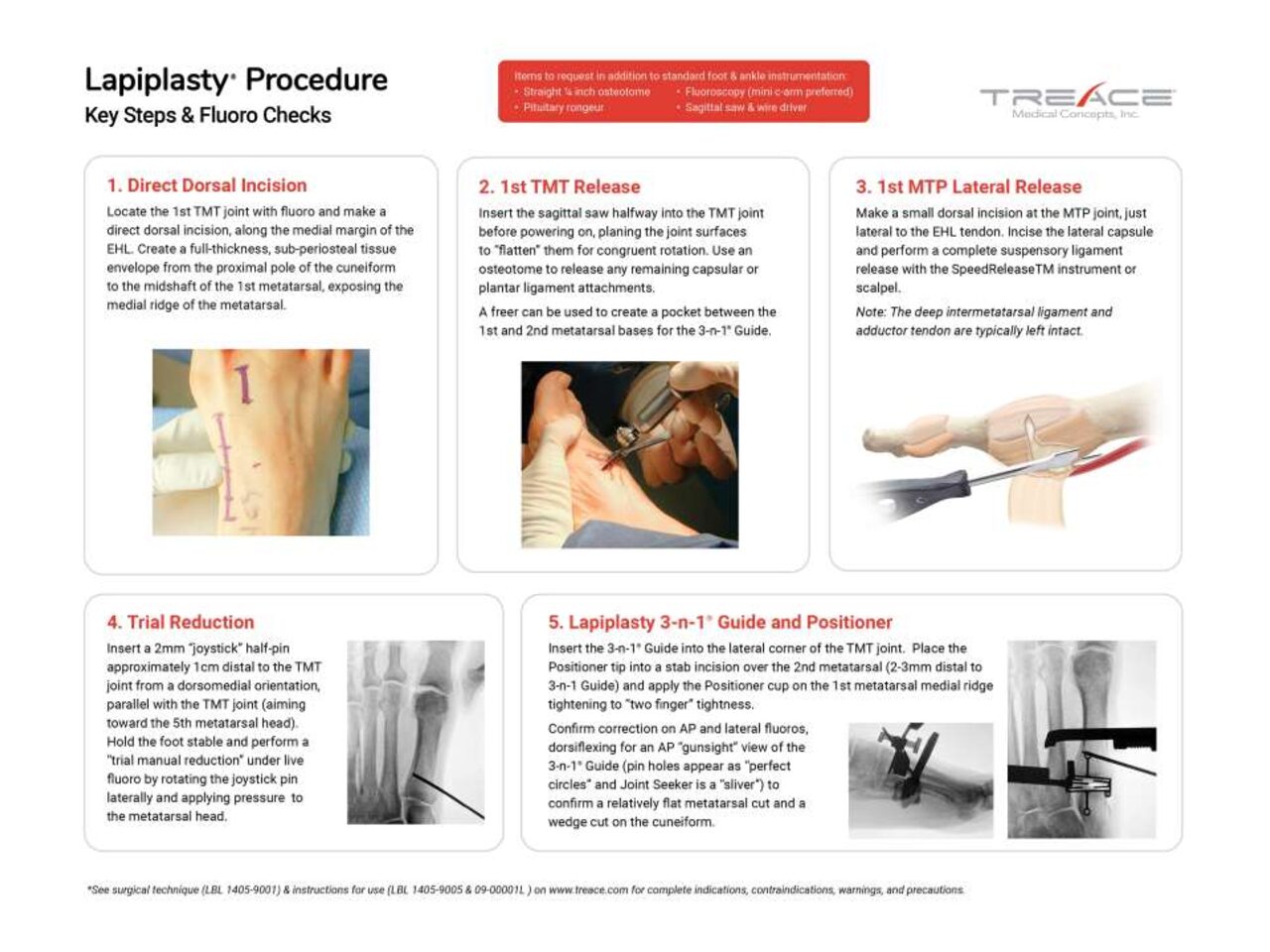
Key Surgical Steps
How the Lapiplasty® Procedure Works

1. Correct
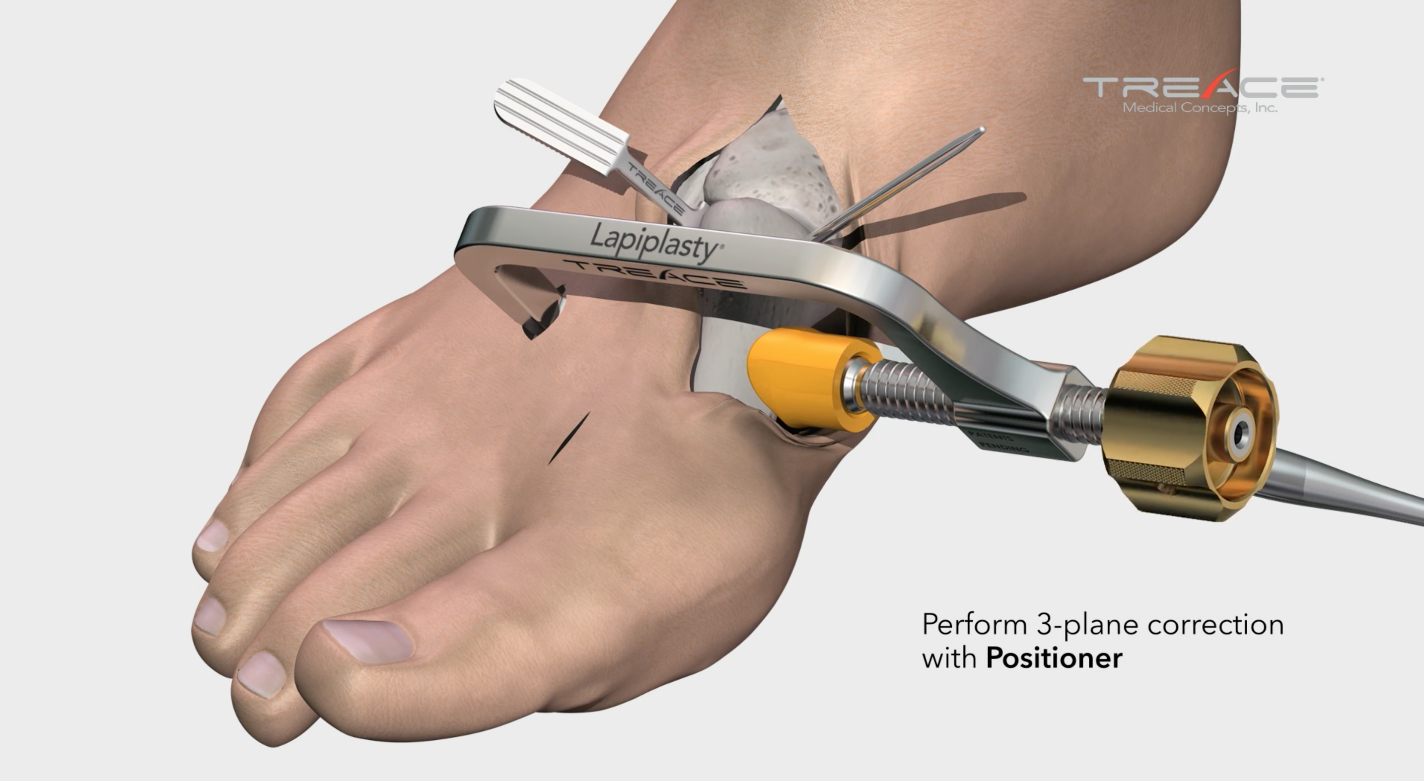
Make Your Correction Before You Cut
The Lapiplasty® Positioner is engineered to quickly and reproducibly correct the alignment in all three planes, establishing and holding true anatomic alignment of the metatarsal and sesamoids.1

2. Cut
Perform Precision Cuts with Confidence
The Lapiplasty® Cut Guide delivers precise cuts with the metatarsal held in the corrected positions, ensuring optimal cut trajectory with only 2.4-3.1mm average metatarsal shortening.3

3. Compress
Achieve Controlled Compression of Joint Surfaces
The Lapiplasty® Compressor is designed to deliver controlled compression4 to the precision-cut joint surfaces, while maintaining 3-plane correction.

4. Fixate
Apply Multiplanar Fixation for Robust Stability
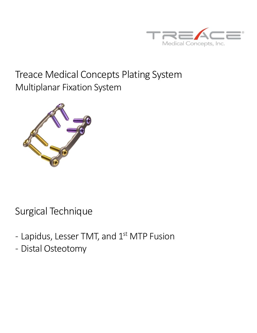
Low-profile Biplanar Plating provides biomechanically-tested5,6 multiplanar stability for rapid return to weight-bearing in a boot.1
Lapiplasty® System
Biomechanically Tested5,6 for Rapid Weight-Bearing1
Our low-profile Biplanar Plating has been biomechanically tested5,6 (and published) to be superior to a standard Lapidus plate and compression screw construct.5

24 Publications and Counting
The Evidence-Based Triplanar Solution
Backed by 24 publications and an ongoing 5-year multicenter prospective study, Treace Medical is recognized as the leader in advancing the scientific study of Hallux Valgus.
Lapiplasty®
3D Bunion Correction®
97 and 99% successful maintenance of 3D correction
(as demonstrated in 13 &17 months follow-up, respectively)1,7
<2 weeks to return to weight-bearing in a boot1,2
10.4mm average reduction in osseous foot width8
2.4 and 3.1mm average shortening of first ray
(in lateral and AP radiographs, respectively)
32-3% non-union rate
(13.5 & 9.5 month follow-up)
1,23% hardware removal rate
(in a 13 month study)
20.9% and 3.2% recurrence rate
(as demonstrated in studies at 17 & 13 months follow-up, respectively)
1,730% increase in cycles to failure with Biplanar Plating
(compared to dorsomedial Lapidus plate + compression screw)
5>80% reduction in pain levels per VAS and MOxFQ scoring systems
(interim analysis from ALIGN3D™ study of 40 patients at 24 months)
9
Now Introducing the Micro-Lapiplasty™ Minimally Invasive System
Now Experience the Power of the Lapiplasty® Procedure through a 2cm Incision
See The Results
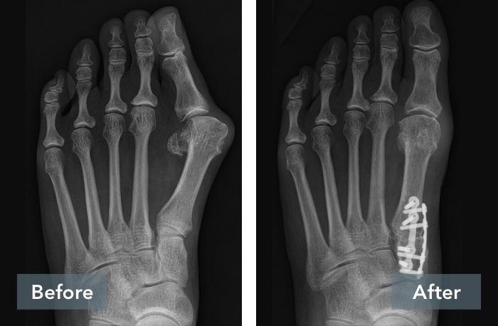
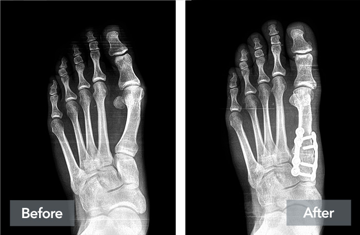
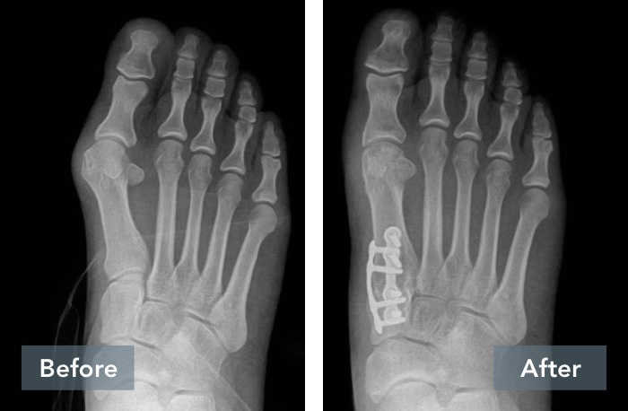
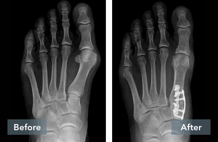
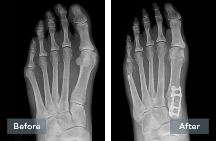
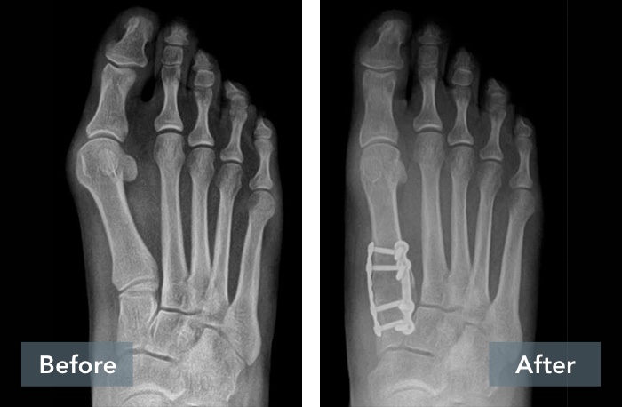
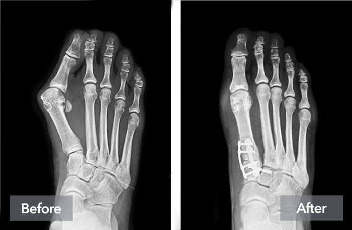
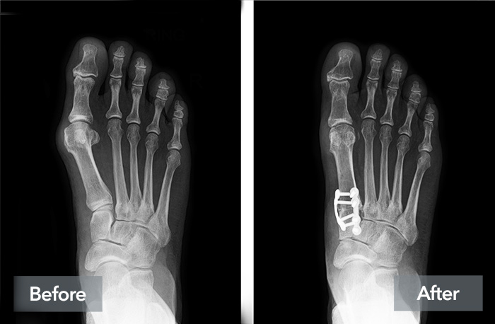
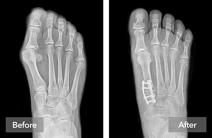
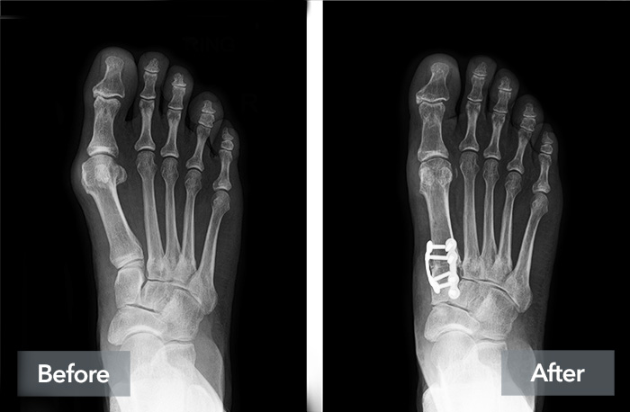
The Beauty of Reproducibility
Scroll through case examples to see the power of Lapiplasty®
Individual results may vary. These experiences are specific to these patients only.
